An HSG is routinely done as part of an initial infertility evaluation and may also be done when investigating possible causes of recurrent miscarriage.
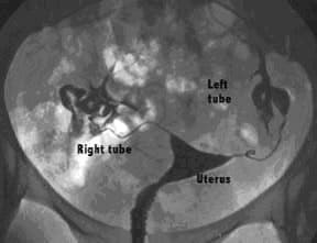
In this procedure, sterile x-ray contrast or dye is introduced into the uterus and flows up into the fallopian tube and out the ends. The dye is visible on x-ray films and therefore the images of the dye reflect the contour of the inside surface of the uterus and tubes. If there is an abnormality of the inside of the uterus (uterine cavity) or blockage of the tubes, this can be detected by examination of the films.
By examining the images of the dye filling the tubes and spilling out the ends, your doctor can predict whether or not the fallopian tubes are open. If the dye does not spill out the end of the tube, this indicates that the tube MAY be blocked. Common causes of tubal blockage include previous infection, endometriosis or previous surgery.
If a “filling defect” (an area where the dye does not fill the uterine cavity) is seen in the uterine cavity, it is possible that a fibroid, polyp or scar tissue is present. A fibroid is a benign smooth muscle tumor of the uterus. Fibroids are very common and present in up to 40% of all women. Usually they do not cause any problems, but if a fibroid protrudes into the uterine cavity, it can cause bleeding, pain, or pregnancy loss. A polyp may also cause a filling defect. Polyps are an overgrowth of the lining inside the uterus. Scar tissue usually results from some sort of trauma to the lining of the uterus, such as an infection or surgical procedure.
The HSG, like any other test, is not 100% effective in detecting any abnormalities. It is generally considered about 90% effective in detecting tubal blockages or filling defects. If abnormalities are detected, other tests may be recommended.
It is important to remember that an HSG usually needs to be scheduled after your period stops, but before ovulation. We recommend that you have someone available to drive you home, and some radiology offices require that someone be available to drive you home. Most patients are able to resume their normal activities almost immediately.
Your doctor may prescribe antibiotics to be taken the day before, the day of, and the day after the procedure. The procedure may occasionally cause mild cramping so it is a good idea to take 600-800 mg of ibuprofen (Motrin/Advil) or 2 Aleve tablets approximately 30-60 minutes prior to your procedure. If these types of medications bother your stomach, take two Extra Strength Tylenol. You can return to your normal activities immediately following your HSG. If you have an allergy to x-ray contrast, iodine or shellfish, or latex, it is very important that you inform us of this at the time of scheduling so that proper preparations may be made for you. Also inform the radiologist of this at the time of the procedure.
The procedure is simple and usually takes about 15 minutes. The following steps are representative of a typical procedure:
After the procedure you may continue to have mild to moderate cramps for several minutes. There may also be some mild cramping throughout the rest of the day. You may also have some light spotting or watery discharge for a day or two. On occasion, there will be a small amount of vaginal bleeding, usually in the form of spotting. You should refrain from intercourse for approximately 48 hours. Other than that, you may immediately resume normal activity. If your cramps are becoming worse instead of better over the course of the day, or if you have any fevers (>100.4F), please call the office.
Advanced Reproductive Medicine & Gynecology of Hawaii
Helping to Create New Beginnings